Developing neuroelectronic hybrids require a deep understanding of the interactions taking place at the cell-device interface. While standard routes involve (bio)chemical functionalization of the surface, our approach emphasizes on biomimetic, low modulus elastic materials as well as topographic features for improving neuron-device interactions. Supported lipid bilayers (SLBs) with embedded cell adhesion and synapse related proteins can be used as biomimetic substrates for neuronal cell culture. A better and more compliant match to the soft brain cells can be obtained by using low modulus elastic materials. Nanotopological cues also play a crucial role in mediating cell attachment, orientation and biocompatibility.
Supported Lipid Bilayers

We are investigating supported lipid bilayer to construct a biomimetic cell-chip interface. Incorporation of positively charged lipid or synaptic adhesion protein has sustained neuronal growth (for more details see papers). Now we are integrating the biofunctionalized surface to our micro/nano structured multi-electrodes.
Soft materials
To investigate the cellular attachment behavior, soft and flexible materials are used. They exhibit a low elastic modulus and are thus very soft, aiming to match the mechanical properties of the brain. Within this area of research, we investigate the influence of the cell on the material and vice versa, looking at whether the cell deforms the substrate or whether the substrate influences the shape of the cell. Furthermore, various polymers of different stiffness are tested.
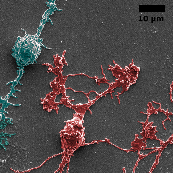
Nanostructured surfaces

Using large area of 3D nanostructured substrates we engage a systematic screening of cellular interactions with respect to the spatial dimensions such as size, distance, and pitch. A special attention is given to cell’s contact with the artificial solid surfaces, and how this surfaces can influence the neurites orientation.
CONTACT:
Prof. Dr. Andreas Offenhäusser
Tel.: +49-2461-61-2330
e-mail: a.offenhaeusser@fz-juelich.de
Dr. Vanessa Maybeck
Tel.: +49-2461-61-3285
e-mail: v.maybeck@fz-juelich.de
Dr. Dirk Mayer
Tel.: +49-2461-61-4023
e-mail: dirk.mayer@fz-juelich.de
PUBLICATIONS:
Ultra‐thin resin embedding method for scanning electron microscopy of individual cells on high and low aspect ratio 3D nanostructures
Belu et al., Journal of microscopy, 2016, 263 (1)
Reconstitution of Fusion Proteins in Supported Lipid Bilayers for the Study of Cell Surface Receptor–Ligand Interactions in Cell–Cell Contact
Ghosh Moulick et al., Langmuir 2016, 32 (14), 3462–3469
How to image cell adhesion on soft polymers?
Seyock et al., Micron, 2017, 92, 39-42